
The PTT dysfunction results in attenuation of these important ligaments and also leads to diminished hindfoot inversion and the peroneus brevis acting unopposed with a dynamic abduction-eversion force. The PTT protects these structures and also plays an essential role in the elastic support of the joint complex 9. The integrity of the talonavicular joint is maintained by the calcaneonavicular ligament (spring ligament) and portions of the superficial deltoid ligament 13. The PTT also works eccentrically throughout the loading response until midstance where the foot is pronated providing controlled lengthening contraction. Therefore, the PTT is critical in inverting the hindfoot and locking the transverse tarsal joint for normal gait and ambulation 13. This enables the gastrocnemius-soleus complex to provide a plantar flexion force against a rigid lever to allow forward progression during the push-off phase of the gait cycle. The divergence at the transverse tarsal joint (calcaneocuboidal and talonavicular joints) allows the subtalar complex to become rigid. The subtalar joint becomes progressively inverted which is initiated by the PTT adducting the transverse tarsal joint. As the foot unloads weight, the resilient arches return to their original shape 2. The plate will untwist, flattening the arches slightly. Immediately after heel strike, the subtalar joint is inverted and the foot is supple 13. When the hindfoot is inverted, the transverse tarsal joint locks and the foot becomes rigid.įigure 1: Twisted osteoligamentous plate of the foot, resulting in longitudinal and transverse arches When the hindfoot is everted the transverse tarsal joint (talonavicular, calcaneocuboid) is unlocked which allows the foot to remain supple. The PTT adducts and supinates the forefoot secondarily inverting the subtalar joint. PTT inserts to 9 bones including navicular tuberosity, 3 cuneiforms, 2nd – 4th metatarsal heads, and sustantaculum tali of calcaneous 2,9. The actual mechanism of twisting and untwisting is accomplished through motion at the talocalcaneonavicular, transverse tarsal, and tarsometatarsal joints that link the bones of the plantar arches 2. The medial longitudinal arch (between the calcaneous and first metatarsal) is higher and more flexible compared to the lateral longitudinal arch (between the calcaneous and lateral metatarsal). The resulting twist forms one transverse and two longitudinal arches (Figure 1).
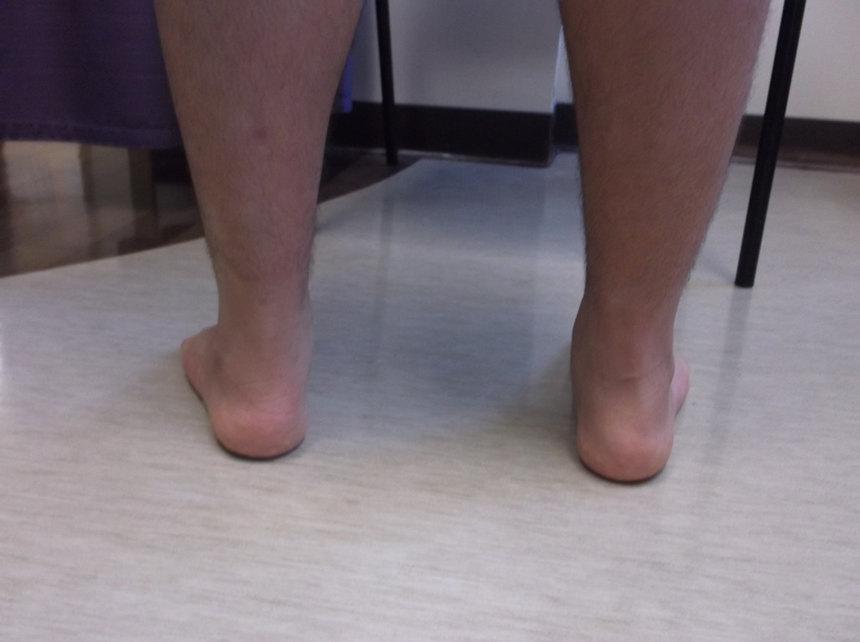
#BILATERAL PES PLANUS FULL#
The anterior edge of the plate (formed by the metatarsal heads) is horizontal and in full contact with the ground and the posterior edge of the plate (the posterior calcaneus) is vertical. The design of the arches can be understood by picturing the foot as a twisted osteoligamentous plate. In addition to these static structures, musculo-tendinous structures (tibialis posterior, peroneal, and tibialis anterior tendons) dynamically stabilize the arch. One transverse and two longitudinal arches are formed by the aligned wedge-shaped tarsal and metatarsal bones, and their corresponding ligaments.

It is most common in females over the age of 40, peaking at 55 years of age, with comorbidities including diabetes and obesity 1,3.Īny injury to the structures mentioned above maintaining the arch may lead to pes planus. These patients develop medial foot pain during late childhood or early adolescence as they increase their physical and sporting activities 3,6.Įpidemiology and Risk factors for PreventionĪdult acquired flatfoot is a debilitating condition that affects up to 5 million people within the US 4. Individuals with congenital pes planus may have tarsal coalition, an abnormal connection between tarsal bones due to incomplete differentiation of bones. Diabetics with Charcot neuroarthropathy from sensory neuropathy commonly have collapsed arches with rocker bottom deformity in severe cases 3.

In severe cases, rupture of PTT occurs commonly at the region of hypovascularity 1.0-1.5 cm distal to the medial malleolus. Dysfunction of the posterior tibial tendon(PTT) leads to pes planus of various degrees. Specific injuries that can lead to pes planus include fractures of the navicular, first metatarsal, or calcaneal bones, and/or trauma to the Lisfranc joint, plantar fascia, and deltoid/spring ligament. In long-standing cases, the ankle may be involved as well 13. It combines multiple static and dynamic deformities, with flattening of the medial arch, eversion of calcaneus, and abduction of forefoot relative to the hindfoot 9. Adult acquired flatfoot deformity (AAFD) is a complex pathology defined by the collapse of the medial longitudinal arch of the foot with continued progressive deformity of the foot and ankle 5.


 0 kommentar(er)
